Home » Integrative Oncology » Page 2

Tag: Integrative Oncology

breast-bone-cancer

The Link between Bone Density and Breast Cancer Risk

Understanding and Monitoring Risk Factors

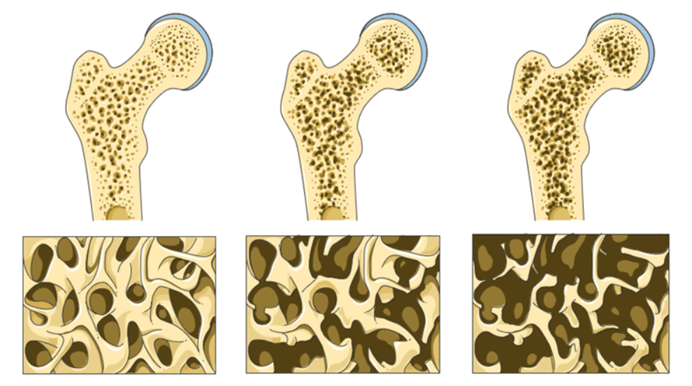
Bone density, or bone mineral density ( BMD ), is the amount of bone mineral in bone tissue.  Bone mineral density (BMD) is a lifetime marker of estrogen exposure in a woman's body and has been associated with increased breast cancer risk. Estrogen is a crucial factor in maintaining bone density and gradually decreases over age. While there are many factors that influence bone density and bone health, the presence of estrogen contributes to the capacity of bone to continuously remodel and maintain the dynamic balance between bone resorption and bone formation.  A woman’s exposure to estrogen over the life cycle may contribute to her risk of breast cancer.

breast-cancer

Bone density measurement is used in clinical medicine as an indirect indicator of osteoporosis and fracture risk.  There is a clear association between poor bone density and a higher probability of fracture.  There is a clear association between poor bone density and low estrogen levels.  Conversely, there is a clear association between increased and healthy bone density and higher estrogen levels.

pink-ribbonScreening for risk of breast cancer should ALSO include assessment of estrogen levels and bone density along with well-recognized risk factors which include first degree relatives, obesity, increased visceral fat, smoking, alcoholism, early menarche, late menopause, sedentary lifestyle, hormone replacement therapy, and prolonged estrogen exposure, increased density of breast tissue. 

I would also add exposure to environmental endocrine disruptors and imbalances in the intestinal microbiome influencing estrogen metabolism.  

Breast density and bone density are related to endogenous and exogenous estrogen exposure in a woman’s body.  There is a correlation between estrogen exposure, high breast density, high bone density, and increased risk of breast cancer.

Bone is living metabolically active tissue. “Bone remodeling is the process by which bone is renewed to maintain bone strength and mineral homeostasis. Remodeling involves continuous removal of discrete packets of old bone, replacement of these packets with newly synthesized proteinaceous matrix, and subsequent mineralization of the matrix to form new bone begins before birth and continues until death.  Bone remodeling increases in perimenopausal and early postmenopausal women and then slows with further aging, but continues at a faster rate than in premenopausal women. Bone remodeling is thought to increase mildly in aging men.”  Normal Bone Anatomy and Physiology 10.2215/CJN.04151206

Engaging in a health model for all patients includes assessing and managing bone health to promote healthy bone over the life cycle. A health model for cancer patients, due to the typically older age demographics will inherently include a large population of patients already at risk for loss of bone mass, osteopenia and osteoporosis. Screening for bone mineral density and managing bone health should be part of whole-person, whole health care. Taking a thorough history that includes family history, bone health and bone mineral density can bring attention to patients at higher risk for low bone density and fracture as well as patients with a higher risk of estrogen driven breast cancers.

Bone density measurement is used in clinical medicine as an indirect indicator of osteoporosis and fracture risk.  There is a clear association between poor bone density and higher probability of fracture.  There is a clear association between poor bone density and low estrogen levels.  Conversely there is a clear association between increased and healthy bone density and higher estrogen levels.

The Most Common Risk Factors for Low Bone Density and Primary Considerations for a Bone Density Test include:

bone-density 

  • Females age 65 or older
  • Males aged 70 or older
  • People over age 50 with
    • previous bone fracture from minor trauma
    • rheumatoid arthritis
    • low body weight
    • a parent with a hip fracture
  • Individuals with vertebral abnormalities
  • Individuals receiving, or planning to receive, long-term glucocorticoid therapy
  • Individuals with primary hyperparathyroidism
  • Individuals being monitored to assess the response or efficacy of an approved osteoporosis drug therapy
  • Individuals receiving androgen deprivation therapy 
  • Individuals with a history of eating disorders

Additional factors that are related to the risk of low bone density and the need for assessment include smoking, alcohol intake, long-term use of corticosteroid drugs, sedentary or convalescent lifestyle, protein status, mineral status, digestion, and absorption function, chronic inflammation and vitamin D status.  

For cancer patients and survivors also consider periods of poor nutrition, calorie, protein status, convalescence, lack of exercise, effect of hormonal therapies, oophorectomy, orchiectomy, chemotherapy, immunotherapy, treatment induced thyroiditis, gastritis, enteritis and colitis,  chronic pain impacting appetite, digestive and absorptive dysfunction, surgical loss of gastrointestinal organs and function as contributors to risk of loss of bone density and as well as multiple and varied adverse effects of cancer physiology and cancer treatments upon nutritional status and active lifestyle.

Selected References 

Clarke B. Normal bone anatomy and physiology. Clin J Am Soc Nephrol. 2008 Nov;3 Suppl 3(Suppl 3):S131-9. doi: 10.2215/CJN.04151206. PMID: 18988698; PMCID: PMC3152283.

Fraenkel M, Novack V, Mizrakli Y, Koretz M, Siris E, Norton L, Shafat T, Geffen DB. Bone mineral density in women newly diagnosed with breast cancer: a prospective cohort study. NPJ Breast Cancer. 2022 Feb 17;8(1):21. doi: 10.1038/s41523-022-00388-z. PMID: 35177701; PMCID: PMC8854387.

Zain NM, Seriramulu VP, Chelliah KK. Bone Mineral Density and Breast Cancer Risk Factors among Premenopausal and Postmenopausal Women A Systematic Review. Asian Pac J Cancer Prev. 2016;17(7):3229-34. PMID: 27509955.

Clinical Pearl-cancer

Clinical Pearl: Chemotherapy Reduces Magnesium to Dangerously Low Levels


Hypomagnesia occurs in 29-100% of cancer patients receiving chemotherapy.

Magnesium deficiency is common in cancer patients, especially those receiving chemotherapy.  Magnesium is the second most abundant intracellular cation after potassium. It is involved in >600 enzymatic reactions in the body.

Hypomagnesia induces  fatigue , mitochondropathy (compromised mitochondrial function )and risk for neuropathy, nephropathy as well as abnormal cardiovascular function (arrhythmia, hypertension) immune dysfunction, headache and altered bone and Vitamin D metabolism.  Hypomagnesia is associated with nausea, vomiting, headache, myalgia, constipation, anxiety, insomnia and depression, all common complaints of cancer patients.

Long term and extreme hypomagnesia promotes cancer treatment related fatigue, cortical blindness, insulin resistance, prolonged QT interval, hypertension, seizures, tremor, psychiatric disturbances, migraine headaches and is associated with chronic inflammation and oxidative stress.

Magnesium status declines with age.

As cancer patients are typically over 50 years old, hypomagnesia may be present long before diagnosis. Pre-menopausal women and athletes also have higher needs of magnesium and may be deficient. 

This may influence the tumor microenvironment towards carcinogenesis, tumorogenesis, proliferation and progression.

Both oral and intravenous repletion relieve many of the hypomagnesia related adverse effects.

Adverse effects can be prevented by supplementing with magnesium in advance of as well as after chemotherapy. In a health model, keep patients replete with Magnesium at times to optimize function, prevent deficiency syndromes and adverse symptoms of chemotherapy.

Monitoring and Management of Magnesium Status

All patient care plans include oral Magnesium Glycinate Chelate

Daily Dose: 600-900mg daily in capsule, liquid or powder form

(Glycinate and Bis-Glycinate chelates are more well absorbed and less likely to have a laxative effect than other forms of magnesium chelate). Excess oral magnesium can lead to diarrhea. Spread out oral dosing over 3-4 doses per day to achieve repletion without loose stool.

Extreme Hypomagnesia can be quickly repleted by intravenous infusion.

All patients are monitored for Serum RBC Magnesium to assess magnesium status every 3-6 months long-term and monthly during active chemotherapy.

Serum Magnesium is not a reliable indicator of Magnesium deficiency.


Dietary Sources of Magnesium include:

Almonds, cashews, brazil nuts, pumpkin seeds, flaxseeds, cocoa, avocados, dark leafy greens, seaweed


Chemotherapeutic agents that induce hypomagnesia:

Platinum Chemotherapy Agents : Oxaliplatin, Cisplatin, Carboplatin and

Taxanes:  paclitaxel (Taxol) nab-paclitaxel (Abraxane), docetaxel (Taxotere),Cabazitaxel (Jevtana).

Vinca alkaloids vinblastine, vincristine, vindesine, and vinorelbine.

Biomarker Lactic Acid Dehydrogenase Predicts Cancer Progression and Overall Survival

Aberrant metabolism and inefficient fuel production are characteristic of tumor cells, which are dominated by aerobic glycolysis, increased lactate production, and a higher uptake of glucose (the Warburg effect). Elevated LDH is a marker of these aberrant metabolic processes in cancer cells. High serum LDH levels are associated with poor prognosis in patients with cancer and predict progression and overall survival.

Aerobic glycolysis was described for the first time about a century ago by Otto H. Warburg who showed that cancer cells metabolize glucose differently than normal cells (Warburg effect) and that tumors derive energy mainly from the conversion of glucose to lactic acid and minimally via cellular respiration involving oxygen. Tumors produce massive amounts of the aerobic glycolysis waste product, lactic acid. This is evidence of deregulated metabolism, hence the understanding of cancer as “disorder of cellular metabolism”.  Lactic Acid itself may promote the growth and spread of cancer cells, especially at high concentrations by changing the tumor microenvironment.

Lactate dehydrogenase (LDH) is an enzyme that catalyzes the reduction of pyruvate to lactate at the end of the glycolytic pathway.

The normal range for LDH is 100-333 u/L, with levels greater than 245 u/L considered to be in the upper quartile of normal.  Elevated LDH, above 245 u/L, is suggestive of early carcinogenesis, tumor cell proliferation, tumor progression, and poor prognosis.

LDH is often highly elevated in aggressive forms of cancer and hematological malignancies including melanoma, lymphoma, acute leukemia, seminoma germ cell, pancreatic, gastric, lung, renal cell, nasopharyngeal, esophageal, cervical, and prostate cancers.

The OutSmart Cancer System® recognizes cancer as a metabolic syndrome and leverages the abnormal metabolism of tumor cells to exert influence over the tumor microenvironment and the behavior of tumor cells. Attending to the Cancer Terrain is a fundamental approach for influencing cancer cell metabolism.  

EGCG, a catechin found in Green Tea (H. Camellia sinensis) has been identified as an agent which inhibits LDH activity in normal and low oxygen environments by influencing the conversion of pyruvate to lactate at the end of the glycolytic pathway.  This may deprive cancer cells of their preferred fuel, glucose, and metabolites, including lactate that produces a favorable environment for malignant proliferation, growth, and progression. Recommended Therapeutic Dose 1-3 grams daily.

Monitoring trends in LDH is a method of both identifying abnormal cellular metabolism found in many solid and hematologic malignancies and is also of value in identifying early signs of recurrence as well as disease progression.

For patients achieving remission, during the first two years after completion of cancer treatment, LDH and other biomarkers of the Cancer Terrain are monitored every 3 months.  Thereafter, every six months for 3-10 more years to track and identify early signs of recurrence.  

For patients living with cancer as a chronic illness, LDH and biomarkers of the Cancer Terrain are monitored every 3 months to track evidence of recurrence and treatment resistance.


Learn more about monitoring the Cancer Terrain and the Tumor Microenvironment.
Receive training in Dr. Nalini’s OUTSMART CANCER SYSTEM ®.
www.aiiore.com

By using biomarkers of the Cancer Terrain and cellular metabolism, it is possible to identify trends that allow for early intervention. LDH is one of the most valuable and reliable biomarkers reflecting the active presence of the aberrant physiology of tumor cells and is prognostic and predictive of progression and overall survival in cancer patients.


Selected References:

Let The Oncologist Be The Disease Expert. Become The Health Expert That Cancer Patients Are Looking For.

You may not treat cancer in your practice, but you do have patients who are at risk due to personal and family history, patients who may be undergoing or recovering from treatments, patients who are survivors worried about recurrence and patients living with cancer as a chronic illness.  And you may also have patients who are family members concerned about their loved ones. 


There is no HEALTH MODEL in conventional oncology care, yet health and wellbeing, peace of mind and sense of agency are in the center of the hearts and minds of cancer patients, cancer survivors and their families. 


There will be 19 million cancer survivors in the US alone by 2024.  Who is supporting their health?  Who is trained to help them recover and keep them well??  …not the oncologist.


How can you help these patients?

A  breast cancer survivor who successfully completed her treatments 8 years ago comes into your office as a new patient complaining of persistent peripheral neuropathy and ongoing cognitive changes since her treatment.  How can you resolve these long-term adverse effects?


An ovarian cancer patient currently undergoing aggressive treatment every 21 days comes into your office complaining of severe diarrhea, neuropathy and sleep disruption.  What can you do to help her get through her treatments with less adverse effects, maintain her weight and nutritional status?


A colorectal cancer survivor who completed his treatment 3 months ago is continuing to have 10-15 bowel movements daily and is profoundly fatigued.  What will you do to restore normal bowel function?


A prostate cancer patient on endocrine blockade therapy is suffering from
hot flashes. Should you also be concerned about loss of bone mass and sleep cycle disruption?


An endometrial cancer survivor is suffering from dermatitis and colitis, adverse effects of her dramatically successful immunotherapy treatment and now has chronic autoimmune inflammation. How will you manage this?


A head and neck cancer patient who has trouble swallowing is losing weight and muscle mass.


How can you provide a plan for repair from oral mucositis, restoration of the oral mircrobiome and repletion of calories and nutrients?


These patients are searching for clinicians that can guide and support them through every phase of their cancer journey.  Just as in helping your patients navigate other chronic illnesses, patients look to you for a plan, for monitoring and guidance so that they can maintain and regain their health during and after their treatments.


When a patient has a collaborative team providing integrative care everyone wins, the patients, families and care providers.  Patients who have a clear plan and support have the opportunity for better outcomes, better prognosis, greater peace of mind, a sense of control and agency and an improved quality of life. 


Let the oncologist be the cancer expert. You can be the health expert on their team.


Standard of care in oncology must  change such that care includes not only a team of disease experts (usually medical oncologist, surgeon, radiologist) but ALSO a team of health experts.


Towards this end  I founded the American Institute of Integrative Oncology Research and Education and  have created an online self-paced training program for front line clinicians who want to expand their skills and their practice and  fill the huge need in our communities and serve these patients.  If you did not specialize in oncology, you probably had one course on this topic but you need to fill the gap in your training to feel confident in doing so.


The Foundations of Integrative Oncology Training is not for clinicians who want to practice oncology.  It is front-line clinicians who want to feel confident, knowledgeable and well trained in supporting the health side of the cancer equation. This self- paced online training is for clinicians who want to increase their impact, expand and grow their practice and represents 35 years of clinical practice and experience.


The first step is learning how to take a comprehensive and complete history of patients whose lives have been touched by cancer.  


You can receive a complimentary copy of the

OUTSMART CANCER CARE PLANNER History and Intake Form

and learn more about the Foundations of Integrative Oncology training here


OutSmart Cancer Care Planner