Home » Omega 3

Tag: Omega 3

fight-prostate-cancer-vegetables

Prostate Cancer Chemoprevention: I3C and DIM

Increasing serum levels of phytochemical DIM may be chemopreventive and chemoprotective for prostate cancer.

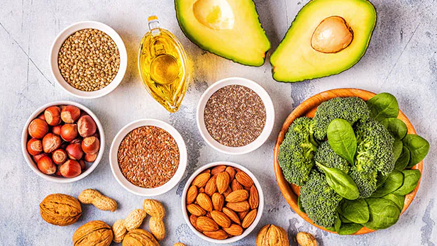
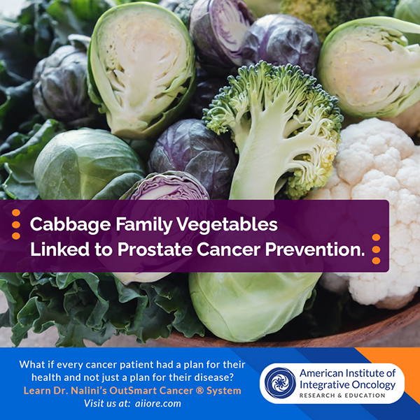
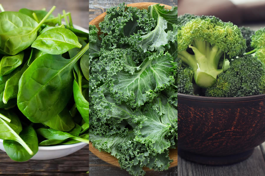
Cruciferous Cabbage Family vegetables such as (including cabbage, cauliflower, Brussels sprouts, broccoli, kale, arugula, bok choy and more ) are rich in dietary phytochemicals including Sulforaphane-Glucosinolate family molecules 13C (Indole-3-Carbinol) and it’s major bioactive therapeutic metabolite DIM  (3 3’di-indole methane).

Chopping, chewing, massaging and lightly steaming cruciferous vegetables activates the plants own catalyzing myrosinase enzyme and exposing the plant to the acid environment in the stomach leading to further metabolism resulting in bio available and bio active DIM.  DIM levels can be measured in the serum.

There are numerous studies on the benefits of dietary consumption of cruciferous vegetables.. A high intake of cruciferous vegetables is associated with reduced risk of several human cancers.  There are strong associations between high intake of broccoli and breast cancer and prostate cancer in humans.  Human cell studies show inhibition of  cell growth of several cancers including breast, prostate, pancreatic, colorectal, lung and head and neck cancers.

DIM and Prostate Cancer

DIM

DIM appears to be a potent inhibitor of human androgen hormones which may promote expression of androgen receptors on prostate cancer cells which may lead to carcinogenesis. Increasing serum levels of DIM through diet and supplementation may therefore be chemopreventive for prostate cancer.

Human and Cell Studies have shown that increased serum levels of DIM 

  • Reduces Serum Prostate Specific Antigen
  • Reduces Serum Androgen Hormones
  • Down Regulates Prostate Stem Cell Activity
  • Decreases  Nuclear Androgen Receptors
  • Induces p450 metabolic detoxification enzymes CYP1A1, CYP1A2 and CYP1B
  • Decreases oxidative stress via nrf2-KEAP pathway

The OutSmart Cancer® System is focused upon transforming the tumor microenvironment  which is a signaling environment, so that there is less physiologic support for the development and spread of cancer.   In a health model (rather than a disease model) we endeavor to transform the biosystem and  reduce pro-carcinogenic and proliferative signaling and prevent cellular, nuclear and mitochondrial damage.

brocolli

Therefore we use phytochemicals such as I3C and DIM and dietary interventions to influence carcinogenic and proliferative signaling in the tumor microenvironment.

Guidelines for Increasing Serum DIM levels

Primary Reference

*Amare DE. Anti-Cancer and Other Biological Effects of a Dietary Compound 3,3ʹ-Diindolylmethane Supplementation: A Systematic Review of Human Clinical Trials. Nutrition and Dietary Supplements. 2020;12:123-137

https://doi.org/10.2147/NDS.S261577

Additional Selected References 

  1. Anderton MJ, Manson MM, Verschoyle RD, et al. Pharmacokinetics and tissue disposition of indole-3-carbinol and its acid condensation products after oral administration to mice. Clin Cancer Res. 2004;10(15):5233–5241. doi:10.1158/1078-0432.CCR-04-0163
  2. Bjeldanes LF, Kim JY, Grose KR, et al. Aromatic hydrocarbon responsiveness-receptor agonists generated from indole-3-carbinol in vitro and in vivo: comparisons with 2, 3, 7, 8-tetrachlorodibenzo-p-dioxin. Proc Natl Acad Sci U S A. 1991;88(21):9543–9547. doi:10.1073/pnas.88.21.9543
  3. Chang Y-C, Riby J, Chang GH-F, et al. Cytostatic and antiestrogenic effects of 2-(indol-3-ylmethyl)-3, 3′-diindolylmethane, a major in vivo product of dietary indole-3-carbinol. Biochem Pharmacol. 1999;58(5):825–834. doi:10.1016/S0006-2952(99)00165-3
  4. Chen I, McDougal A, Wang F, et al. Aryl hydrocarbon receptor-mediated antiestrogenic and antitumorigenic activity of diindolylmethane. Carcinogenesis. 1998;19(9):1631–1639. doi:10.1093/carcin/19.9.1631
  5. Bradfield CA, Bjeldanes LF. High-performance liquid chromatographic analysis of anticarcinogenic indoles in Brassica oleracea. J Agric Food Chem. 1987;35(1):46–49. doi:10.1021/jf00073a010
  6. Weng J-R, Tsai C-H, Kulp SK, et al. Indole-3-carbinol as a chemopreventive and anti-cancer agent. Cancer Lett. 2008;262(2):153–163. doi:10.1016/j.canlet.2008.01.033
  7. Bradlow HL. Indole-3-carbinol as a chemoprotective agent in breast and prostate cancer. In Vivo. 2008;22(4):441–445
  8. Ahmad A, Ali S, Wang Z, et al. 3, 3′‐diindolylmethane enhances taxotere‐induced growth inhibition of breast cancer cells through downregulation of FoxM1. Int J Cancer. 2011;129(7):1781–1791. doi:10.1002/ijc.25839
  9. Ali S, Banerjee S, Ahmad A, et al. Apoptosis-inducing effect of erlotinib is potentiated by 3,3ʹ-diindolylmethane in vitro and in vivo using an orthotopic model of pancreatic cancer. Mol Cancer Ther. 2008;7(6):1708–1719. doi:10.1158/1535-7163.MCT-08-0354
  10. Banerjee S, Wang Z, Kong D, et al. 3, 3′-Diindolylmethane enhances chemosensitivity of multiple chemotherapeutic agents in pancreatic cancer. Cancer Res. 2009;69(13):5592–5600. doi:10.1158/0008-5472.CAN-09-0838
  11. Giovannucci E, Rimm EB, Liu Y, et al. A prospective study of cruciferous vegetables and prostate cancer. Cancer Epidemiol Prev Biomarkers. 2003;12(12):1403–1409.
  12. Kong D, Heath E, Chen W, et al. Loss of let-7 up-regulates EZH2 in prostate cancer consistent with the acquisition of cancer stem cell signatures that are attenuated by BR-DIM. PLoS One. 2012;7(3):e33729. doi:10.1371/journal.pone.0033729
  13. Abdelbaqi K,Lack N, Guns ET, et al. Antiandrogenic and growth inhibitory effects of ring‐substituted analogs of 3, 3′‐diindolylmethane (Ring‐DIMs) in hormone‐responsive LNCaP human prostate cancer cells. Prostate. 2011;71(13):1401–1412.
  14. Bhattacharjee S, Dashwood RH. Epigenetic Regulation of NRF2/KEAP1 by Phytochemicals. Antioxidants. 2020; 9(9):865. https://doi.org/10.3390/antiox9090865
  15. Li Y et al. Recent progress on nutraceutical research in prostate cancer. Cancer Metastasis Rev. 2014;33(2-3):629-640.
omega 3 fatty acids

Omega 3 Fatty Acids: Enhanced Control of Cancer Risk and Progression

A diet high in polyunsaturated fatty acids, especially omega 3s, have been shown to be negatively associated with cancer development

 Dietary fatty acids have been recognized as influential factors in the activation of carcinogenic events or disease progression and have been associated with a direct connection to breast cancer prevention.

PUFAs differentially inhibit mammary tumor development by inflicting modifications to the morphology of cell membranes, and influencing signaling pathways, gene expression and apoptosis.

The human body is unable to synthesize long-chain polyunsaturated fatty acids (PUFAs) Omega 3 DHA, docosahexaenoic, and EPA, Eicosapentaenoic acid and Omega 6 Arachidonic Acid at a reasonable rate and therefore, supplementation is required through dietary sources or nutritional supplements. The recommended daily nutritional dose is 2,000 mg EPA+DHA, while therapeutic dosing is 4,000-6,000 milligrams of EPA+DHA per day.

omega-3-natural

 Omega Three Fatty Acids and the Tumor Microenvironment

  1. Supports Normal Inflammation Control by lowering COX 2, LOX5, PGE2, IL1, IL6,TNFa, CRP.
    • Increased inflammation contributes to cancer development, progression and metastasis.
    • Increased inflammation is linked to cancer related pain, fatigue, depression and cognitive impairment.
    • Increased inflammation is linked to cancer related hypercoagulation and risk of thromboembolism
    • Supporting Normal Inflammation control has a wide impact on the behavior of tumor cells and on safety and quality of life for cancer patients and survivors.
  2. Promotes Expression of M1 Type Tumor Associated Macrophages (TAMs).
    • Type M1 TAMs promote tumor regression, inflammation control and immune activation by promoting tumor infiltration by antigen presenting dendritic cells and cytotoxic T cells.
  3. Inhibits VEGF (Vascular Endothelial Growth Factor) and Promotes Normal Control of Angiogenesis .
    • VEGF promotes the development of new blood vessels to the tumor cells. Inhibition of VEGF and the development of capillaries inhibits tumor growth and profession as well as metastasis.
       
  4. Down regulates tumor promoter Protein Kinase C isoenzymes,
    • A group of enzymes that link multiple cellular processes responsible for regulation of tumorigenesis, cell cycle progression and metastasis.
  5. Inhibits Collagenase,
    • A proteolytic enzyme that breaks down the ECM (Extracellular Matrix) and allows invasion of tumor cells into tissues and blood vessels, leading to progression, invasion and metastasis.
  6. Promotes Normal Apoptosis signaling.
    • Cancer cells lose the ability to initiate apoptosis, the normal process in which a cell recognizes itself as aberrant and self destructs. The inhibition of normal apoptotic signaling in malignant cells is a hallmark  of the tumor microenvironment permissive of uncontrolled growth, persistence and immortality due to loss of normal regulation.
  7. Lowers Bcl2 and Ras oncogenes.
    • These genes inhibit normal apoptosis and promote tumor growth and progression.
  8. Acts as a Chemo-sensitizer
    • Working synergistically to enhance therapeutic effect of chemotherapy drugs. DHA has a potential to specifically chemo-sensitize tumors.
    • Tumour cells can be made more sensitive to chemotherapy than non-tumor cell when membrane lipids are enriched with DHA
    • Incorporating DHA during treatment reduces adverse effects of chemotherapy.
    • DHA can improve the outcome of chemotherapy when highly incorporated into cell membranes.
  9. Acts as a Radio-sensitizer.
    • By promoting normal membrane structure and function and by influencing the tumor microenvironment DHA acts synergistically to potentiate therapeutic effects of radiotherapy on tumor cells.
  10. Promotes Healthy 16-OH Estrogen metabolism.
    • Estrogen can be metabolized through multiple pathways. The promotion of 16-Hydroxylation of estrogen produces estrogen metabolites that are not pro-carcinogenic. Omega 3 Fatty Acids promote healthy estrogen metabolism.
  11. Inhibits Platelet Aggregation and Thrombin Formation.
    • Abnormal hyper-coagulation, increased platelet aggregation and thrombus formation are hallmarks of the tumor microenvironment. Control of platelet aggregation and thrombus formation reduces the risk of life threatening and adverse  thrombotic events.  40% of all cancer patients are at risk for the formation of thromboembolisms.  Omega 3 Fatty Acids reduce this risk.
  12. Promotes Normal Cell Membrane Functions and Receptor Binding
    • A healthy flexible cell membrane built of omega 3 fatty acids promotes an enhancement of all membrane functions, normalizing and optimizing normal and therapeutic physiology.
  13. Increases expression of Tumor Suppressor Gene PTEN.
    • Increased expression of tumor suppressor genes leads to enhanced control over carcinogenesis,  tumorigenesis and metastatic progression.
  14. Inhibits Multi Drug Resistance.
    • Tumor cells can quickly become resistant to therapeutic anti-neoplastic agents thus decreasing and shortening the efficacy of treatments.
  15. Inhibits cachexia preserves muscle mass and bone mass (inhibits proteolysis inducing factor)
    • Loss of bone mass (osteopenia) and loss of muscle mass (sarcopenia) are risk factors of aging and of the cancer physiology.  Maintaining bone mass and muscle mass are crucial to robust healthy function and quality of life.
  16. Supports normal mood regulation.
    • Depression and anxiety are common in cancer patients. Support of balanced mood allows cancer patients deep and restful sleep, improved quality of life and increased coping capacity and resilience in the face of stress.

Cautions and Contraindications

  • Patient on anticoagulant medications
  • Patients with thrombocytopenia and known hypo-coagultion clotting disorders
  • Pre and Post Surgical patients (72 hours)
  • Patients with seafood allergies


How to Measure Omega 3 Fatty Acid Status

Serum or Plasma Omega 3 Fatty Acid ratios. LABCORP Omega 3-6 Fatty Acids, Quest Diagnostics Omegacheck, Boston HeartLab Fatty Acid Balance, Cleveland HeartLab Omegacheck, Genova Diagnostics Essential and Metabolic Fatty Acids, Great Plains Comprehensive Fatty Acids, OmegaQuant Omega3 Index.

Selected References

 Azrad M, Turgeon C, Demark-Wahnefried W. Current evidence linking polyunsaturated Fatty acids with cancer risk and progressionFront Oncol. (2013) 3:224.

 Bartsch H, Nair J, Owen RW. Dietary polyunsaturated fatty acids and cancers of the breast and colorectum: emerging evidence for their role as risk modifiers. Carcinogenesis. (1999) 20:2209–18.

 Bournoux, P. Et al. Improving outcome of chemotherapy of metastatic breast cancer by DHA: Phase II Trial, Br.J Cancer 2009 Dec 15:101(12):1978-85

 Shweta Tiwary   Altered Lipid Tumor Environment and Its Potential Effects on NKT Cell Function and Tumor Immunity.  Front Immunol.10.3389/fimmu.2019.02187

 Zanoaga O, Jurj A, Raduly L, Cojocneanu-Petric R, Fuentes-Mattei E, Wu O, et al. Implications of dietary omega-3 and omega-6 polyunsaturated fatty acids in breast cancer.  Exp Ther Med. (2018) 15:1167–76. 10.3892/etm.2017.5515